
Congratulations! You have finally entered into the awe-inspiring and transformative journey that will bring your miracle into this world. During your pregnancy, your doctor will guide you to help you take better care of yourself as well as your baby. What more is involved in this prenatal care, are the ultrasounds which will provide you some glimpses of the happenings inside your womb as your baby develops and grows.
The goal of prenatal care is to ensure your safety as well as your baby’s well being. Ultrasounds are a great way to keep track of the baby’s development as well as know or predict any problems that may occur. A gynecologist in Karachi described in detail the importance of ultrasound visits with your doctor during pregnancy and what happens in each one. Let us have a look.
What Is A Pregnancy Ultrasound?
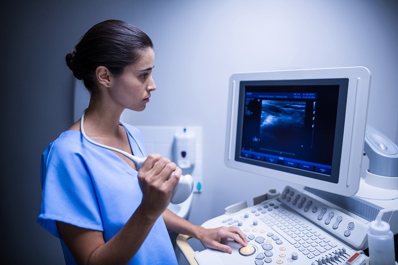
Getting a sneak peak of your bundle of joy is by far the most beautiful moment of this journey as per the claim of the majority of pregnant women. An ultrasound, commonly known as a sonogram, is a way to create and see the images of your internal organs. It is usually performed by your OB-GYN during your pregnancy. The ultrasound uses sound waves to create images of your baby inside the womb. What is important to mention here is that these sound waves are not harmful for your baby and are considered safe.
The ultrasound creates sound waves, which come into contact with body tissues and organs and are then bounced back. These waves bounce back as echoes to the equipment which then turn it into an image called a sonogram. This may be available in various dimensions.
Types Of Ultrasounds
There are majorly two main types of ultrasounds that are done during your pregnancy.
Transvaginal ultrasound: This ultrasound is usually done in the early stages of the pregnancy. This provides a more accurate view of the baby as compared to the other one in the early stages. A transducer is inserted into the vagina to view the baby. It is then moved around for better visuals.
Transabdominal ultrasound: This is done by moving a transducer over the abdomen (baby bump) to get clear images of the baby’s anatomy and movements. You may lay on your back and the OB-GYN moves the transducer ave your abdomen after applying a thin layer of gel over your bump. This gel allows the sound waves to move easily.
Ultrasounds In The First Trimester
During the first trimester, the baby’s organs have not developed to the point where we can differentiate them. This is usually done to estimate the gestational age i.e. how long the pregnancy has progressed and what would be the possible due date. This is also used to screen the baby for any genetic disorders. It is entirely up to you if you want to do genetic screening at this stage as people have various opinions on said matter. Some women prefer to have as much information as possible while others do not.
This is the point where it will be possible to see whether the mother will be having a single baby or twins, or even more. Fetal heart rate is also checked in these ultrasounds. Another important thing to note is the location of the implant where the fetus is going. At this point, it will be detected whether the pregnancy is in the uterus or if it is an ectopic pregnancy. An ectopic pregnancy is one where the fertilized egg grows out of the uterus. Unfortunately, this pregnancy is not a viable one and does not last to term.
Ultrasounds In The Second Trimester
Usually done between 18 to 22 weeks of pregnancy, this is the time when the baby has developed to the point where organs can be differentiated. The baby’s limbs and their development will be checked. It is also known as an anatomy scan. The baby’s gender is also determined at this stage.
At this point, the doctor will look for any abnormalities in the brain, heart, liver , kidneys, and other vital organs. The heart’s function is checked in quite detail. Amniotic fluid levels and the location of the placenta is also seen via these ultrasounds.
Ultrasounds in Third Trimester
The baby’s growth and positioning as well as the location of the placenta is seen in ultrasounds that are done in the third and last trimester. Many fetal abnormalities and growth restriction issues can be detected in these. Many people do not get a sonogram in the third trimester. Those with risks of fetal developmental issues or other complications are recommended to get ultrasounds done in the third trimesters.
Final Thoughts
These are just a fascinating way to get to know your baby’s developmental progress during pregnancy. It is better to get those done as and when the doctor sees fit to avoid any unwanted complications later. Better to get to know of issues beforehand rather than later.